Friday 23 November 2012
Thursday 22 November 2012
Gastrointestinal stromal tumours (GISTs)
GISTs are rare connective tissue tumours that show similar differentiation patterns to the interstitial cells of Cajal and approximately 60e70% arise in the stomach.
Activating mutations of the KIT or PDGFRA protooncogenes are thought to be the causal molecular events.
Approximately 95% of tumours are therefore positive for CD117 (c-KIT) by immunohistochemistry.
The degree of local and metastatic spread of gastric GISTs should be evaluated by CT scan and endoscopic ultrasound.
If the tumour is localised to the stomach it should be surgically resected.
If the tumour is unresectable or if metastases are present at diagnosis or during follow-up, treatment with the tyrosine kinase inhibitor, imatinib, should be considered and treatment should usually be continued until progression, intolerance or patient refusal occurs.
‘Consensus meeting for the management of gastrointestinal stromal tumors’.


Tuesday 20 November 2012
Liver diseases in pregnancy
1- Hyperemesis Gravidarum:
1-20/1000 in the 1st trimester, 50% hospitalized
25% have abnormal liver biochemistry
raised conjugated bilirubin, ALP x2 normal, ALT up to 200iu/l
aetiology unknown
? rapidly rising steroid level
complications related to repeated vomiting
outcome of pregnancy normal
2-per-eclampsia:
hypertension, protienurea, odema
5-7% of pregnancies
2nd and 3rd trimester
can lead to seizures, renal failure, coma and death
hepatic infarction and rupture occur rarely
3-HELLP syndrome:
complication of severe pre-eclampsia
4-20% eclamptic pregnancies
3rd trimester 2/3, postpartum 1/3
Haemolysis (elevated LDH)
ELevated liver enzymes (ALT x 2-10)
Low Platelets (<100 p="">presentation: N/V, malaise, headache, RUQ pain, hepatomegaly.
mortality: 1% maternal, 35% fetal
severe morbidity: DIC, placental rupture, renal failure, pul odema.
lab tests: uric acid >7.8, LDH >600, ALT>100, smear: haemolysis
4-AFLP acute fatty liver of pregnancy:
rare, 1:7000-13000, potentially fatal
3rd trimester, 1st and multiple pregnanies
Mild features of pre eclampsia
Jaundice after 1-2 weeks
hypoglycaemia, clotting abnormality
Untreated leads to fulminant hepatic failure
lab tests: elevated ALT and bilirubin, DIC, coagulopathy, elevated amonia, renal failure
maternal mortality up to 10-20%, fetal mortality 20-50%
5-obstetric cholestasis:
2nd and 3rd trimester (>26 weeks)
prurius (plams and soles)
juandice uncommon 25%
raised ALT 60%, gamma GT 30%
raised serum bile acids
imaging of the lliver normal
close monitoring, delivary by 37-38 weeks
treat with UDCA and/or dexamethsone, vit K prevent post-partum bleed
The serum-ascites albumin gradient
SAAG is a calculation used in medicine to help determine the cause of ascites.
SAAG = (albumin concentration of serum) - (albumin concentration of ascitic fluid).
High gradient
A high gradient (> 1.1 g/dL) indicates the ascites is due to portal hypertension with 97% accuracy.
Important causes of high SAAG ascites (> 1.1 g/dL) include:
high protein (> 2.5): heart failure, Budd Chiari syndrome
low protein (< 2.5): cirrhosis of the liver, nephrotic syndrome
Low gradient
A low gradient (< 1.1 g/dL ) indicates causes of ascites not associated with increased portal pressure such as tuberculosis, pancreatitis, nephrotic syndrome and various types of peritoneal cancer.
A low gradient (< 1.1 g/dL ) indicates causes of ascites not associated with increased portal pressure such as tuberculosis, pancreatitis, nephrotic syndrome and various types of peritoneal cancer.
King's college criteria
The most widely accepted prognostic tool for patients who present with ALF. Although fulfilment of these criteria has a high specificity for mortality, the sensitivity and negative predictive value remain low. Therefore, not fulfilling the criteria does not ensure survival.
Prospective data from the US Acute Liver Failure Study Group revealed the King's College Criteria had an overall specificity of 85.7% but lower sensitivity of 48.3% in prediction of mortality in 838 patients with ALF. The specificity was increased to 92.4% in the 374 patients who presented with ALF secondary to paracetamol (acetaminophen) overdose.
ALF secondary to paracetamol overdose:
-pH <7>
-INR >6.5 (PT >100 seconds) and
-serum creatinine >300 micromol/L (>3.4 mg/dL) in patients with grade 3 or 4 hepatic encephalopathy.
-pH <7>
-INR >6.5 (PT >100 seconds) and
-serum creatinine >300 micromol/L (>3.4 mg/dL) in patients with grade 3 or 4 hepatic encephalopathy.
Non-paracetamol associated ALF:
INR > 6.5 (PT > 100 sec) or
any 3 of the following:
-age between 10-40 yrs,
-aetiology non-A, non-B hepatitis or idiosyncratic drug reaction,
-duration of juandice before hepatic enchephalopathy > 7days,
-INR >3.5 (PT >50),
-Serum bilirubin > 300 mmole/l

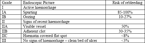
Forrest classification
Used for stratifying patients with upper gastrointestinal hemorrhage into high and low risk categories formortality. It is also a significant method of prediction of the risk of rebleeding and very often is used for evaluation of the endoscopic intervention modalities.

Rockall score
Identify patients at risk of adverse outcome following acute upper gastrointestinal bleeding.
The scoring system uses clinical criteria (increasing age, co-morbidity, shock) as well as endoscopic finding (diagnosis, stigmata of acute bleeding).
A convenient memnoic is ABCDE - i.e. Age, Blood pressure fall (shock), Co-morbidity, Diagnosis and Evidence of bleeding.

The utility of a modified Rockall score (that is, a score lacking endoscopic findings) has not been established.
The definition of mild, moderate, or severe risk remains a matter of clinical judgement.
According to SIGN guidleines, only patients with Rokall score of 0 can be savely managed as outpatient.
The predicted mortality:
Score 0 0.2%
Score 1 2.4%
Score 2 5.6%
Score 7 50%
MELD score
Scoring system for assessing the severity of chronic liver disease.
It was initially developed to predict death within three months of surgery in patients who had undergone TIPS and was subsequently found to be useful in determining prognosis and prioritizing for receipt of a liver transplant.
This score is now used by the United Network for Organ Sharing (UNOS) and Eurotransplant for prioritizing allocation of liver transplants instead of the older Child-Pugh score.
The MELD range from 6 to 40 points. This range predict 3 months survival without liver transplantation in patients with liver cirrhosis.MELD = 3.78[Ln serum bilirubin (mg/dL)] + 11.2[Ln INR] + 9.57[Ln serum creatinine (mg/dL)] + 6.43
- 40 or more — 71.3% mortality
- 30–39 — 52.6% mortality
- 20–29 — 19.6% mortality
- 10–19 — 6.0% mortality
- < 9 — 1.9% mortality
The United Kingdom MELD (UKELD) score is derived from the patient's serum sodium, creatinine and bilirubin, and INR.
Minimal listing criteria require that the patient should have a projected 1-year liver disease mortality without transplantation of more than 9%. This is predicted by a UKELD score of 49 or greater.
A UKELD score of 60 is predictive of a 50% 1-year survival.
at MELD score of 15 or greater, survival is enhanced 1 year after liver transplantation versus remaining in the waiting list. Therfore, most transplant centres designate a MELS score of 15 as the minimal listing score for liver transplantion.
Child-Pugh score
Used in patients with liver cirrhosis to assess the severity of the clinical condition.
Five variables are considered (severity of ascites and of encephalopathy, abnormality in the serum bilirubin, serum albumin and clotting times), and a score (of between 1 and 3) is accordingly assigned to each of these factors. This grade is used as a general means to verify the prognosis of the patient.
Child A 1 yr survival: 100%, 2 yr survival:85%
Child B 1 yr survival: 81%, 2 yr survival:57%
Child C 1 yr survival:45%, 2 yr survival:35%
 |
| Child-Turcotte-Pugh classification, My Gastro Room blog |
Monday 19 November 2012
Crohn's disease Vs ulcerative colitis

Mucosal architecture:
Mucosal surface, normal, irregular, villous
Crypt atrophy (shortened, widely spaced crypts)
Distorted, dilated, branching crypts
Inflammatory changes:
Basal plasmacytosis, increase in cells in basal third of lamina
propria
Increased lamina propria cellularity (round cells and
neutrophils)
Basal lymphoid aggregates
Specific features:
Epithelioid granuloma
Basal giant cells
Excess histiocytes in lamina propria
Features related with activity (separating IBD from normal):
Neutrophils in surface epithelium
Neutrophils in crypt epithelium
Ulceration
UC:
Architecture:
Severe crypt architectural distortion
Severe widespread decreased crypt density
Frankly villous surface
Inflammatory:
Heavy diffuse transmucosal lamina propria cell increase
Diffuse basal plasmacytosis
Miscellaneous:
Increased intensity of the alterations towards the distal colon
Severe mucin depletion
Paneth-cell metaplasia distal to the hepatic flexure
CT:Non-Cancerous cystic Liver Lesions


Focal nodular hyperplasia is the second most common liver mass.
Appears as lobulated intrahepati lesions with central lucency (scar)

a large, round, low attenuated lesion with enhancing ring.
Rt lobe abscess + Rt hemidiaphragm elevation + recent travel =Amebic liver abscess
aspiration is not mandatory.

Typical echinococcal liver cyst in the right lobe of the liver.
sheepherder + cough + liver cyst with septations, eggshell calcification and hydated sand =Echincoccosis
 |
| Haptic adenoma |
Well demarcated mass with early enhancement in the arterial phase before iso-attenuation in the delayed phase. strongly associated with OCP. MRI spoked wheel appearance. Can rupture: resect/ embolise.
Crohn's Disease

Wilson disease

Noalcoholic fatty liver disease

Mallory's hyaline bodies (pink filamentous structures,black arrowhead) are cytoplasmic inclusions in hepatocytes consisting of abnormal keratin, hyaline, and other proteins. They are usually found in hepatocytes that are ballooned (black arrow) and are morphologic hallmarks of alcoholic and nonalcoholic steatohepatitis. Mallory's bodies are not cause but rather consequence of cellular injury. Usually hepatocytes with Mallory's bodies do not contain large fat vacuoles, although microvesicular fat may be seen. In this frame, other hepatocytes are present, containing macrovesicular fat globules (white arrow), which occupy almost all cytoplasm, displacing nucleus (white arrowhead) to periphery

alpha-1-antitrypsin deficiency

It has been stained with period-acid Schiff, which strongly stains intrahepatic globules pink [long black arrow]. The hepatocytes containing the globules are concentrated adjacent to a band of fibrous tissue [long black and white arrow]. A mild inflammatory infiltrate and myofibroblastic cells are seen associated with the fibrous tissue [short black and white arrow]. It is likely that this patient has early fibrosis wild mild active hepatitis. If this continues the patient is at risk of developing overt cirrhosis.
Sunday 18 November 2012
Cystic Pancreatic tumours
1- Serous Cystadenoma:
10% of all cystic lesions of the pancreas
Large, palpable asymptomatic upper abdominal mass.
occur equaly in males and females
CT scan: shows a lobulated cystic mass with a central scar, microcytic cluster of small cysts. 20-30% have stellate scar "sunburst".
EUS: shows multiple anechoic cystic cavities
Histologically consist of multiple tiny cysts that contain watery fluid.
While SCAs rarely have malignant potential, surgical resection for symptomatic lesions or lesions greater than 4 cm is recommended in good operative candidates.

2- Mucinous Cystadenoma:
Almost exclusively in women 40-60 years old
Usually have thick fibrotic walls and can be multiple
Cysts do not communicate with the main pancreatic duct
1-2% of pancreatic endocrine tumours
Histologically consist of multiple cysts containing sticky mucous
The stroma is refered to as 'ovarian like'
Treatment is surgical resection in approperiate patients.

3-Intraductal Papillary Mucinous Tumour IPMT:
Consist of intraductal papillary growth of mucin-producing columnar epithelium.
obstructive pancreatits frequently occurs
CT: dilated main or side ducts
Main duct IPMN: Because of the high risk of invasive cancer in the main duct, surgical resection is recommended ( Whipple's )
Side chain IPMN: still high risk of malignancy. advice resection or survay
At ERCP, half of the patients have the diagnostic finding of a patulous papilla extruding mucous.

coeliac disease

typical duodenal scalloping that is seen in patient with celiac sprue
Marsh classification:
- Marsh stage 0: normal mucosa
- Marsh stage 1: increased number of intra-epithelial lymphocytes, usually exceeding 20 per 100 enterocytes
- Marsh stage 2: proliferation of the crypts of Lieberkuhn
- Marsh stage 3: partial or complete villous atrophy
- Marsh stage 4: hypoplasia of the small bowel architecture
esophageal candidiasis

esophageal candidiasis can be confirmed with esophageal brushings.
The best treatment option for candida esophagitis is a systemic antifungal agent such as fluconazole.
Topical antifungal agents such as nystatin may not sufficient.
eosinophilic esophagitis (EoE)
eosinophilic esophagitis (EoE) :
 |
| Esinophilic oesophagitis ( Dr E Said) |
The esophageal rings in this patient with eosinophilic esophagitis (EoE) represent more esophageal fibrosis with remodeling than inflammation.
Remember that endoscopy in EoE is unreliable and often confusing. A history of dysphagia is a clear indication for biopsy sampling even in the absence of evident abnormalities.
Represent a chronic, immune mediated eosophageal disease characterised clinically by symptoms related to oesophageal dysfunction and histologically by an eosinophilic predominent inflammation.
EoE is food related disease that persumed to result from eosionphilic activation to deitary antigen which is limited to the oesophagus.
? abnormalities in chromosome 7
Symptoms:
Adults:
1-Dysphagia for solids 100%
2-Long lasting food impaction 35%
3-Non swallowing related retro sternal pain 50%
Children:
1-Food refusal
2-Failure to thrive
3-Vomiting, regurgitation
4-Chest pain/ abdominal pain
5-Diarrhea
Histology:
> 15 eso/ hpf
eosinophils micro abscesses
surface laying of eosinophils
Its a patchy disease, so several Bx should be taken from proximal and distal oesophagus.
Treatment:
1-Diet:
elemental/ elemination diet
6 food diet elimination ( caw milk, wheat, eggs, soy, nuts,seafood, shellfish )
2-Drugs:
topical corticosteroids
3-Dilatation:
post procedure pain, risk of deep ulceration/ perforation
pseudomembranous colitis
Pseudomembranous colitis is due to C. difficile infection, gram-positive bacillus that colonize the gut following transmission via the feco-oral route and disruption of the gut flora following course of antibiotic.
The organism produces 2 exotoxins: toxin A (enterotoxin) and toxin B (cytotoxin).
C. difficile May be detected in 1-3% of healthy adults and up to 50% of infants and children carry it.
Treatment of asymptomatic carriers is not recommended.
Pseudomembranous colitis occurs in only 10% of cases of antibiotic-associated diarrhea.
In 5-19%, it may be localized to the proximal colon.
Studies suggest that C. difficile and inflammatory bowel disease are being seen together more frequently. Rates have increased 2-fold for Crohn’s disease and 3-fold for ulcerative colitis.
When severe disease is encountered as manifested by pseudomembranes, colon wall thickening, WBC>15,000, creatinine rise by 50%, decreased albumin, and/or increased lactate treatment with vancomycin 125 mg qid for 7-14 days is recommended.
Recurrent C. difficile infection is thought to occur in 10-25% of patients. After one recurrence, additional recurrences are 40-60% more likely.

Autoimmune pancreatitis
AIP is type of chronic pancreatitis characterized by an autoimmune inflammatory process in which prominent lymphocye infiltration with associated fibrosis of the pancreas cuases organ dysfunction.
AIP is one component of multisystem IgG4 associated fibro-inflammatory disease.
The diagnosis is based on combination of clinical, radiological and pathological criteria.
Clinical features include:
-painless jaundice
-upper abdominal discomfort
-Wt loss
-DM i in 75%
-other associaed autoimmune disorders
Radiological features:
-CT: sausage-shaped pancreas
-ERCP: low CBD stricture, diffuse pancreatic stricturing.
histologicaly, there are 2 types:
1-Lymphoplasmacytic LPSP
IgG4 positive,
fibrosis > inflammation
older age, M > F
2-Idiopathic duct centric IDCP
IgG4 negative ( i.e no serum marker)
inflammation > fibrosis
younger age, M=F
Treatment:
corticosteroids
The failure to differentiate AIP from malignancy may lead to unnecessary pancreatic resection.

sausage-shaped pancreas
The CT image displays the characteristic pattern of autoimmune pancreatitis. The pancreas is "sausage" shaped and has a characteristic inflammatory "halo." The initial mode of therapy is oral prednisone which is effective in over 60-70% of cases.
The CT image displays the characteristic pattern of autoimmune pancreatitis. The pancreas is "sausage" shaped and has a characteristic inflammatory "halo." The initial mode of therapy is oral prednisone which is effective in over 60-70% of cases.
Saturday 17 November 2012
Acute Pancreatitis

|
|
Intestinal resection and short bowel syndrome
Intestinal resection is usually well tolerated, but massive resection is followed by the short-bowel syndrome.
The effects of resection depend on the extent and the areas involved.
Because the gut is long, a 30-50% resection can usually be tolerated without undue problems.
Shortened bowel depends on several factors:
-the length of the bowel resected
-the location of the bowel resected
-the integrity of the bowel remaining
-the presence of the colon
Residual jejunum shows less capacity for structural and functional adaptation than residual ileum.

Note:
if less than 100 cm if the ileum is resected, the liver can compansate for the loss of absorptive capacity by producing an increased amount of bile salts, which enter the colon and cause bile irritant diarrhea. this type of diarrhea is treated with cholestyramine, which bind the excess bile salts and improves diarrhea.note:
The effects of resection depend on the extent and the areas involved.
Because the gut is long, a 30-50% resection can usually be tolerated without undue problems.
Shortened bowel depends on several factors:
-the length of the bowel resected
-the location of the bowel resected
-the integrity of the bowel remaining
-the presence of the colon
Residual jejunum shows less capacity for structural and functional adaptation than residual ileum.
| The ileum has specific receptors for the absorption of bile salts and vitamin B12, so that relatively small resections will lead to malabsorption of these substances. Removal of the ileocaecal valve increases the incidence of diarrhoea. The following occur in ileal resection:
small bowel follow-through, measurement of B12, bile salts and occasionally fat absorption. hydrogen breath test will show rapid transit. Many patients require B12 replacement and some need a low-fat diet if there is steatorrhoea. If diarrhoea is a problem, colestyramine, which binds bile salts, often helps. |
| Jejunal resection: |
| The ileum can take over the jejunal absorptive function. Jejunal resection may lead to gastric hypersecretion with high gastrin levels; the exact mechanism of this is unclear. Structural and functional intestinal adaptation take place over the course of a year, with an increase in the absorption per unit length of bowel. |
| Massive intestinal resection (short-bowel syndrome): |
| This most often occurs following resection for Crohn's disease, mesenteric vessel occlusion, radiation enteritis or trauma. There are two types of short-bowel syndrome:  |
| 1-Shortened small intestine ending at a terminal stoma: |
| The major problem is of sodium and fluid depletion and the majority of patients with 100 cm or less of jejunum remaining will require parenteral supplements of fluid and electrolytes, often with nutrients. Sodium losses can be minimized by increasing salt intake, restricting clear fluids between meals and administering oral glucose-electrolyte mixture with a sodium concentration > 90 mmol/L. Jejunal transit time can be increased and stomal effluent loss of fluids and electrolytes reduced by treatment with the somatostatin analogue octreotide, and to a much lesser extent, with loperamide, codeine phosphate or co-phenotrope. There is no benefit of a low-fat diet, but fat assimilation can be increased on treatment with colestyramine and synthetic bile acids. |
| 2-Shortened small intestine in continuity with colon |
| Only a small proportion of these patients require parenteral supplementation of fluid, electrolytes and nutrients because of the absorptive capacity of the colon for fluid and electrolytes. Unabsorbed fat results in impairment of colonic fluid and electrolyte absorption so patients should be on a low-fat diet. A high carbohydrate intake is advised as unabsorbed carbohydrate is metabolized anaerobically to short-chain fatty acids (SCFAs). SCFAs are absorbed and act as an energy source (1.6 kcal/g) and stimulate fluid and electrolyte absorption in the colon. Patients are often treated with colestyramine, which binds dihydroxy bile acids which otherwise have a deleterious effect on colonic fluid and electrolyte absorption and increase colonic oxalate absorption to form renal stones. |

if less than 100 cm if the ileum is resected, the liver can compansate for the loss of absorptive capacity by producing an increased amount of bile salts, which enter the colon and cause bile irritant diarrhea. this type of diarrhea is treated with cholestyramine, which bind the excess bile salts and improves diarrhea.note:
if more than 100 cm is resected, the liver can no longer compansate. the resulting bile salt deficiency leads to steatrrhea.this can be managed by prescribing diet that consist of medium chain triglycerides.
Bacterial overgrowth
| The gut contains many resident bacteria with anaerobic bacteria, e.g. Bacteroides, bifidobacteria, being 100-1000 times more abundant than aerobic (facultative anaerobes), e.g. Escherichia, Enterobacter, Enterococcus. This gut microflora has major functions: -metabolic, e.g. fermentation of non-digestible dietary residues into short chain fatty acids as an energy source in the colon. -initiate vitamin K production. -control epithelial cell proliferation -involved in the development and maintenance of the immune system. -protect the gut mucosa from colonization by pathogenic bacteria. |
| The upper part of the small intestine is almost sterile, containing only a few organisms derived fromthe mouth. Gastric acid kills most organisms and intestinal motility keeps the jejunum empty. The normal terminal ileum contains faecal-type organisms, mainly Escherichia coli and anaerobes and the colon has abundant bacteria |
| Bacterial overgrowth is normally only found associated with a structural abnormality of the small intestine, although it can occur occasionally in the elderly without such abnormality. E. coli and/or Bacteroides, both in concentrations greater than 106/mL are found as part of a mixed flora. These bacteria are capable of deconjugating and dehydroxylating bile salts, so that unconjugated and dehydroxylated bile salts can be detected in aspirates by chromatography. The clinical features are chiefly diarrhoea and steatorrhoea. Steatorrhoea occurs as a result of conjugated bile salt deficiency. |
| The bacteria are able to metabolize vitamin B12 and interfere with its binding to intrinsic factor, thereby leading to B12 deficiency. Conversely some bacteria produce folic acid giving a high serum folate. |
| Bacterial overgrowth has only minimal effects on other substances absorbed from the small intestine. The vitamin B12 deficiency is not so severe as to produce a neurological deficit. Confirmation of bacterial overgrowth is with the hydrogen breath test; aspiration studies are not routinely performed. |
| Although bacterial overgrowth may be responsible for the presenting symptoms, it must be remembered that many of the symptoms may be due to the underlying small bowel pathology. |
| Treatment |
| If possible, the underlying lesion should be corrected (e.g. a stricture should be resected). With multiple diverticula, grossly dilated bowel, or in Crohn's disease, this may not be possible and rotating courses of antibiotics are necessary, such as metronidazole, a tetracycline, or ciprofloxacin. |
Subscribe to:
Posts (Atom)